Pelvic Anatomy Dog : Male Anatomy And Histology - The pelvis is firmly attached to the spine (sacroiliac joint) and the limb is longer and more angulated than the thoracic limb (which is designed to bear weight and absorb impact).
Pelvic Anatomy Dog : Male Anatomy And Histology - The pelvis is firmly attached to the spine (sacroiliac joint) and the limb is longer and more angulated than the thoracic limb (which is designed to bear weight and absorb impact).. Secure the dog's pelvic limbs in this position using tape around the femurs at the level of the stifle joint (figures 2a and 2b). They are most commonly seen in young, healthy dogs and cats subsequent to being hit by car. The canine ischiatic or ischial tuberosities are wide and project caudally to form a broad ischiatic table. This is a really nice video tour of the hip and pelvis, and even though it talks about the human hip, the anatomy is very similar in the dog. Muscles of the pelvic limb.
The institute of canine biology: Muscles of the pelvic limb. The canine pelvis is positioned between the dorsal and transverse planes and closer to the dorsal plane. This veterinary anatomical atlas includes selected labeling structures to help student to understand and discover animal anatomy (skeleton, bones, muscles, joints, viscera, respiratory system. It provides information about a dog's skeletal, reproductive, internal, and external anatomy, along with accompanying labeled diagrams.
Bryan Gross Anatomy Pelvic Cavity Flashcards Cram Com from images.cram.com Providing a larger stride for. Secure the dog's pelvic limbs in this position using tape around the femurs at the level of the stifle joint (figures 2a and 2b). Transect the lateral head of the gastrocnemius near its origin and reflect it off of the underlying digital flexor m. A non articular depression portion of the acetabulum used for the attachment of the ligament of the head of the femur. This is a really nice video tour of the hip and pelvis, and even though it talks about the human hip, the anatomy is very similar in the dog. Has cranial and caudal bellies in the dog only. Anatomy is a branch of biology and medicine that studies the morphology and structure of living organisms. The canine pelvis is positioned between the dorsal and transverse planes and closer to the dorsal plane.
Concurrent injuries to vital organs are very common, and should be addressed before definitive fracture management.
The girdle musculature and the rump muscles. The size of hindlimb bones varies due to the significant variation in size for. However, dogs don't have a collar bone, unlike humans; K eep reading to learn more!. Muscles of the pelvic limb. You will see that there are both ligaments and muscles that play important roles in stabilizing the hip joint, and developing the strength in these muscles through appropriate exercise is important for a. The pelvis must fracture in three places, if displacement is to occur. A non articular depression portion of the acetabulum used for the attachment of the ligament of the head of the femur. It provides information about a dog's skeletal, reproductive, internal, and external anatomy, along with accompanying labeled diagrams. The canine pelvis is positioned between the dorsal and transverse planes and closer to the dorsal plane. The canine pelvis is relatively small and narrow. Dog anatomy is not very difficult to understand if a labeled diagram is present to provide a graphic illustration of the same. Miller's anatomy of the dog, ed 4, st loius, 2013, saunders/elsevier.)
In 94% sacral and in 87.3% medial il … The muscles and nerves of the canine pelvic limb are reviewed, including muscle actions.if you find this helpful, please let me know by like it. Pelvis anatomy bones joints ligaments and foramina kenhub Anatomy_of_pelvis_in_dogs 3/3 anatomy of pelvis in dogs anatomy of pelvis in dogs yeah, reviewing a books anatomy of pelvis in dogs could grow your near friends listings. This veterinary anatomical atlas includes selected labeling structures to help student to understand and discover animal anatomy (skeleton, bones, muscles, joints, viscera, respiratory system.
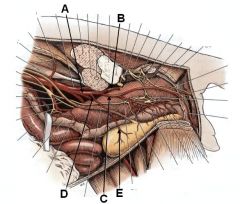
2 from The girdle musculature and the rump muscles. Transect the lateral head of the gastrocnemius near its origin and reflect it off of the underlying digital flexor m. The pelvic girdle is formed by two hip bones which are joined ventrally at the cartilagenous pelvic symphysis and articulate dorsally with the sacrum. Providing a larger stride for. When properly aligned, the patella for each pelvic limb will be centered within the trochlear groove over the distal femur. Anatomy_of_pelvis_in_dogs 4/8 anatomy of pelvis in dogs section on exotics covers the anatomy of ferrets, rodents, rabbits, snakes and lizards in addition to the anatomy of dogs, cats, horses, pigs, cows, goats, and birds. Urogenital system of the dog the pictures in this section are reprinted with permission by the copyright owner, hill's pet nutrition , from the atlas of veterinary clinical anatomy. The term pelvic bladder involves displacement of the bladder from its normal position and affected size and/or position of the urethra.
The canine pelvis is relatively small and narrow.
Anatomy of the dog illustrated atlas this modules of vet anatomy provides a basic foundation in animal anatomy for students of veterinary medicine. Although the internal iliac arterysupplies the pelvis with branches that are named the same in the dog and cat, the branching pattern is different in the two species. And female dog anatomy aims at making a study of all parts of the female dog's body. K eep reading to learn more!. This veterinary anatomical atlas includes selected labeling structures to help student to understand and discover animal anatomy (skeleton, bones, muscles, joints, viscera, respiratory system. However, dogs don't have a collar bone, unlike humans; Dog anatomy is not very difficult to understand if a labeled diagram is present to provide a graphic illustration of the same. Concurrent injuries to vital organs are very common, and should be addressed before definitive fracture management. The bone that articulates with the hip bones to form the hip joint is the femur. With the large range of breeds and dog sizes, despite their difference in appearance, it might be surprising to hear dog anatomy is generally the same with regards to physical anatomy and characteristics. In 243 mongrel female dogs anatomy, topography of the pelvic lymph nodes (ln), composition and frequency of their revealing have been studied. This is a really nice video tour of the hip and pelvis, and even though it talks about the human hip, the anatomy is very similar in the dog. The canine ischiatic or ischial tuberosities are wide and project caudally to form a broad ischiatic table.
The girdle musculature and the rump muscles. Types and functions of dog anatomy Medial aspect of the genual region. Dog anatomy comprises the anatomical studies of the visible parts of the body of a domestic dog.details of structures vary tremendously from breed to breed, more than in any other animal species, wild or domesticated, as dogs are highly variable in height and weight. The pelvis is firmly attached to the spine (sacroiliac joint) and the limb is longer and more angulated than the thoracic limb (which is designed to bear weight and absorb impact).
3 from The canine pelvis is positioned between the dorsal and transverse planes and closer to the dorsal plane. The muscles and nerves of the canine pelvic limb are reviewed, including muscle actions.if you find this helpful, please let me know by like it. It has the ability to flex extend rotate adduct and abduct its whole limb because of this. Medial aspect of the genual region. This veterinary anatomical atlas includes selected labeling structures to help student to understand and discover animal anatomy (skeleton, bones, muscles, joints, viscera, respiratory system. The hindlimb skeleton of the canine includes the pelvic girdle, consisting of the fused ilium, ischium, and pubis, and the bones of the hindlimb. By means of roentgenological and morphological methods in 100% of cases, lateral, iliac and hypogastric ln are revealed. The institute of canine biology:
Dogs have a skeletal system.
Muscles of the pelvic limb; Medial aspect of the genual region. The canine pelvis is relatively small and narrow. K eep reading to learn more!. The size of hindlimb bones varies due to the significant variation in size for. Transect the lateral head of the gastrocnemius near its origin and reflect it off of the underlying digital flexor m. Urogenital system of the dog the pictures in this section are reprinted with permission by the copyright owner, hill's pet nutrition , from the atlas of veterinary clinical anatomy. Dog anatomy is not very difficult to understand if a labeled diagram is present to provide a graphic illustration of the same. Concurrent injuries to vital organs are very common, and should be addressed before definitive fracture management. However, dogs don't have a collar bone, unlike humans; Muscles of the pelvic limb. This is a really nice video tour of the hip and pelvis, and even though it talks about the human hip, the anatomy is very similar in the dog. They are most commonly seen in young, healthy dogs and cats subsequent to being hit by car.
You will see that there are both ligaments and muscles that play important roles in stabilizing the hip joint, and developing the strength in these muscles through appropriate exercise is important for a pelvic anatomy. As understood, exploit does not recommend that you have fantastic points.


0 Komentar